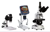
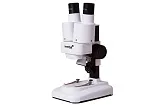
Step-by-step guide on microscope slides preparation and further use
What can be more fun than exploring this diverse colorful microscopic world using your own independently prepared microscope slides! Some people prefer to acquire ready-to-use microscope slides in shops specializing in such things. Of course, that’s easier and will save you a little time. And some of our customers are simply not aware of how to do it and in what sequence. That’s why we are about to share with you all required information about this delicate, but truly interesting process of preparing microscope slides at home! After you read this article this task won’t cause any difficulties anymore. Moreover, you will be able to observe all those interesting things that can be found around your own house! So let’s begin!
You will need:
- Flat blank glass slide
- Glass slide with cavity
- Pipette
- Cover slip
Microscope slide preparation using flat slide glass
1. Make sure that the blank slide,
cover slip and dropper that you are going to use are perfectly clean, dry and dust free.
2. Place the microscope glass slide on a clean, dry surface.
3. Use your pipette to draw a few droplets of the studied liquid.
4. Gently squeeze the dropper so that a single drop falls in the center of the microscope glass slide.
5. Accurately lower the cover slip on the drop. First, lower one of its edges and then the rest of it. Do not press down on the cover slip after you have placed it on the glass slide.
6. Take this ready-to-use microscope slide and carefully place it on the microscope stage.
Microscope slide preparation using concavity slide
1. Using the pipette, draw a few drops of the liquid you want to examine.
2. Take the concavity glass slide by holding it by the edges with two fingers.
3. Gently squeeze three or four drops of liquid, using the pipette, into the slide’s concave depression, or well.
4. Place the slide on the microscope stage.
Tips and warnings
- A blank slide is a flat piece of glass.
- A concavity slide or cavity slide is designed to hold a few drops of liquids.
- Always hold your glass microscope slides and cover slips by their edges. Touching other parts may cause fingerprints on their surfaces which negatively affect image quality.
A typical fresh liquid microscope specimen “lives” about 10-30 minutes before it dries out. This time can be extended by up to several days by applying a thin layer of petroleum jelly on all four sides of the cover slip and then covering it in the conventional manner. The next thing to do is to apply light pressure on the edges of the cover slip. This procedure will create something like a sealed container and will prevent the sample from drying out too quickly.
How to study glass microscope slides
Observing microorganisms under a microscope is a very simple and interesting activity! The procedure is quite simple: The microscope slide should be placed on a microscope stage and then clamped with special spring clips. But viewing slides under a microscope requires careful attention in order to increase the educational effect and minimize the chance of breakage.
The detailed steps:
1. Put your microscope on a flat surface. If your microscope is equipped with an electric illuminator, make sure that you have an outlet nearby.
2. To carry your microscope from one place to another, keep one hand on the tube holder, and the other hand under the base of the tripod. This will allow you to avoid accidental drops and breakage of the instrument.
3. If your microscope is equipped with an illuminator, connect the microscope to the power source to turn it on, and then remove the eyepiece cover.
4. Holding the microscope slide by the edges, place it on the microscope stage (the cover slip should be on top). Slowly move the microscope slide so the specimen is in the center right under the microscope objective lens.
5. Turn the revolving nosepiece so that the objective lens with the smallest increase (usually the smallest in size as well) is imposed on the specimen. Using the coarse adjustment knobs, lower the objective lens as closely as possible to the slide, but without making contact with the slide. Control the process looking directly at the microscope stage (not through the eyepiece). This is especially important when working with high-powered objectives.
6. Looking through the eyepiece, slowly lift the objective lens away from the specimen, using the coarse adjustment knobs, until the image is in focus. After that, use the fine adjustment mechanism to obtain the maximum sharpness of the image.
7. Change the objective lens to a more powerful one by turning the revolving nosepiece. Remember to control the distance between the objective lens and the stage to avoid damage to the objective or specimen. Repeat step 5 - this time with a higher magnification power. Use this method every time you change magnification of the instrument.
One piece of advice before you start
Although objective lenses with low magnification never come too close to the slide, it is necessary to visually check the distance between the objective lens and the slide. High-power objectives can be easily lowered below the permissible range. This will damage the specimen or objective lens. It’s crucial to develop good habits while working with the instrument to prevent accidental damage of delicate optical elements.
Any reproduction of the material for public publication in any information medium and in any format is prohibited. You can refer to this article with active link to levenhukb2b.com.
The manufacturer reserves the right to make changes to the pricing, product range and specifications or discontinue products without prior notice.


